
Please contact the tool managers directly.
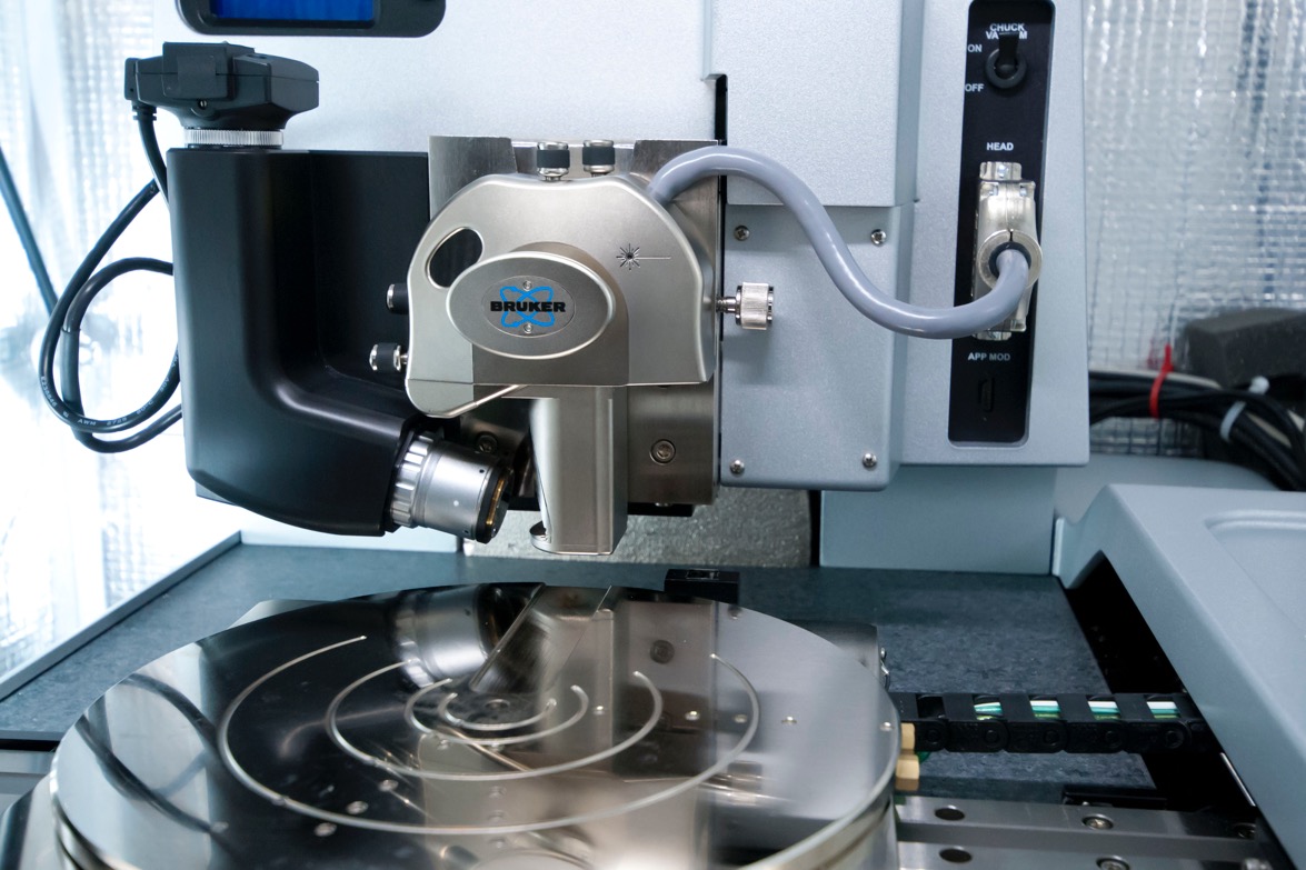
- 6 wafers or smaller
- Magnetic pucks available for small pieces
The operational principle of an atomic force microscope is described by considering a surface of interest being scanned with a sharp tip residing at the free end of a microfabricated cantilever beam. The apex of the tip either gently contacts the surface when imaging is performed in contact mode, or intermittently contacts the surface during tapping mode imaging. The ultrasmall repulsive or attractive forces existing between the tip and the sample cause the cantilever to move up and down in the direction vertical to the surface. This deflection is monitored using an optical deflection setup consisting of a laser beam focused at the free end of the oscillator reflecting into a quad cell photodetector. During this scanning process, bending deflection, oscillation and torsion of the cantilever can be simultaneously measured
In a similar manner as in tapping mode AFM, where the cantilever oscillation is driven at resonance utilizing a piezoelectric driver, phase imaging utilizes an additional signal of the bimorph excitation. In this imaging mode, the phase lag between the cantilever oscillations relative to the signal of the excitation is monitored. The phase lag is extremely sensitive to variations in the adhesion forces and viscoelasticity of the surface. The dynamics of the tip-surface interaction are generally described by assuming a hertzian contact wherein essential deformation forces are described by an elastic sphere pressed into an elastic surface. Due to the tip-surface coupling, this description predicts both a shift of the resonant frequency as well as phase. Frequency shifts are generally not used since ambient operating conditions of the AFM contribute to considerable air damping of the oscillator producing quality factors less than 10. Alternatively, phase shift imaging is employed in imaging multicomponent surfaces by mapping local variations of mechanical properties which are absent in height imaging.
- Metrology sub-Angstrom resolution
- 100x100 µm lateral dynamic range
- 10 µm vertical dynamic range
- Tapping Mode
- Contact Mode
- ScanAsyst Mode
- NanoMan